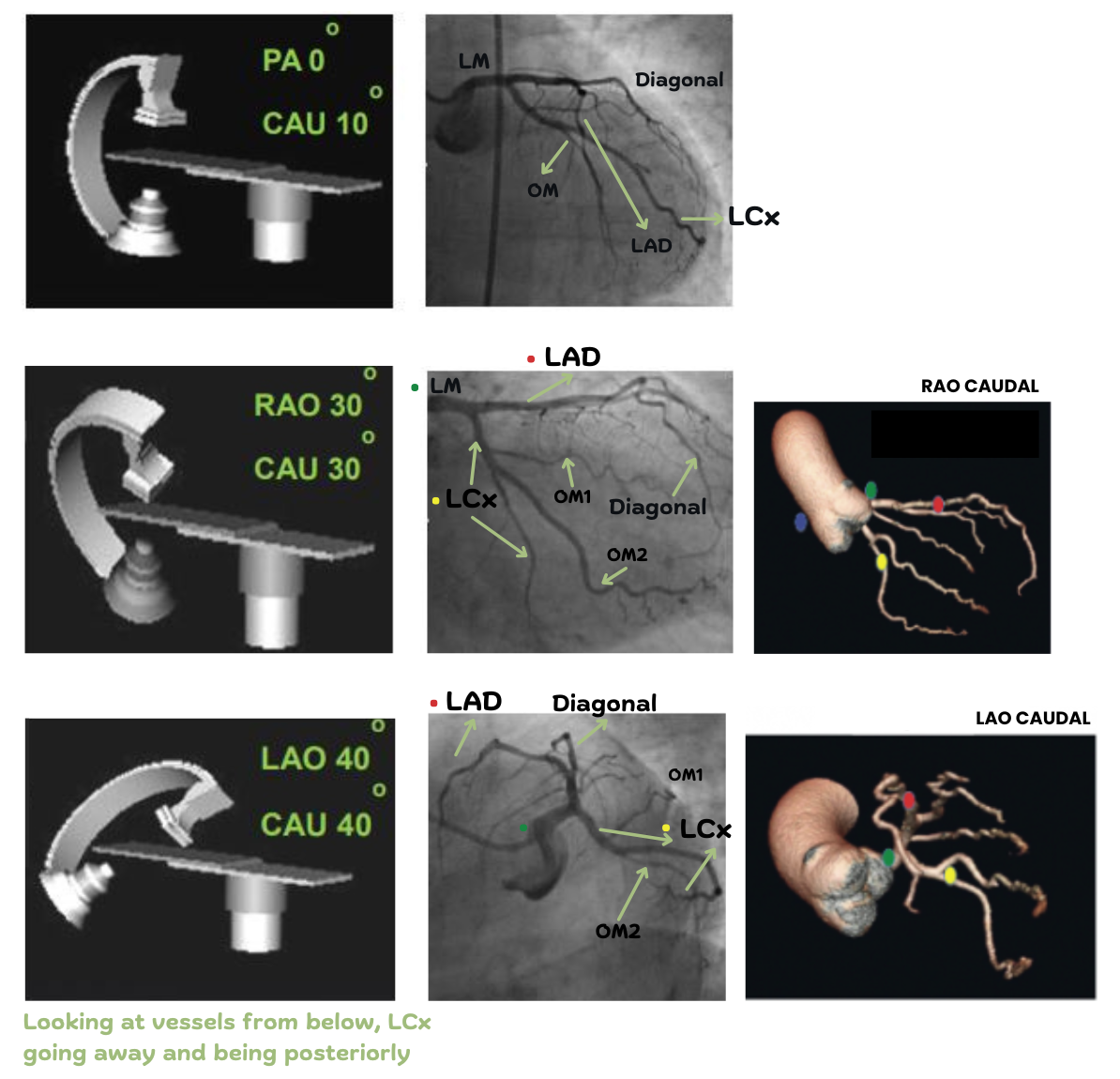
Left Coronary Arteries
Typically, 5 views are used to visualize the LCA and the 2 main branches:
- LAD (Left anterior descending) Diagonals and Septals
- LCx (Left circumflex) Obtuse Marginals (OM)
LAD
Supplies anterior portion of LV (critical region) - usually imaged/checked first if possible.
♥ Cranial views elongate the LAD so it is shown running down towards the apex of the heart
- Diagonals Branch off to the left
- Septals Branch off to the right
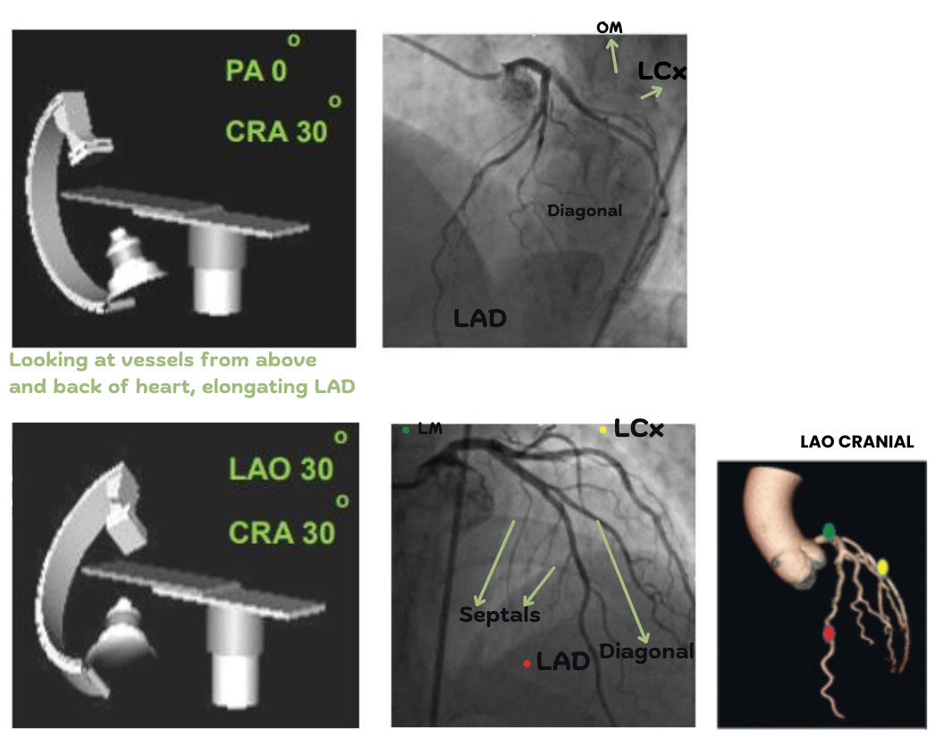
LCx
Supplies left atrium and postero-lateral aspect of LV.
♥ Caudal views show the LCx running down the back of the heart.
The last image is called the 'spider view', which shows the ostium or beginning of the major vessels bifurcating from the LM (left main) - good for visualising proximal stenosis.
- OM Branch off down
Normal Variants
- Due to stenosis, smaller vessels may expand to compensate (e.g. septals appearing larger than the LAD)
- There may be collateral connections between the left and right sides to compensate for stenosis
- (LCx) If the OM comes off the LM directly, it is called a ramus instead
- (LM) Some people have a very short LM that bifucrates almost immediately
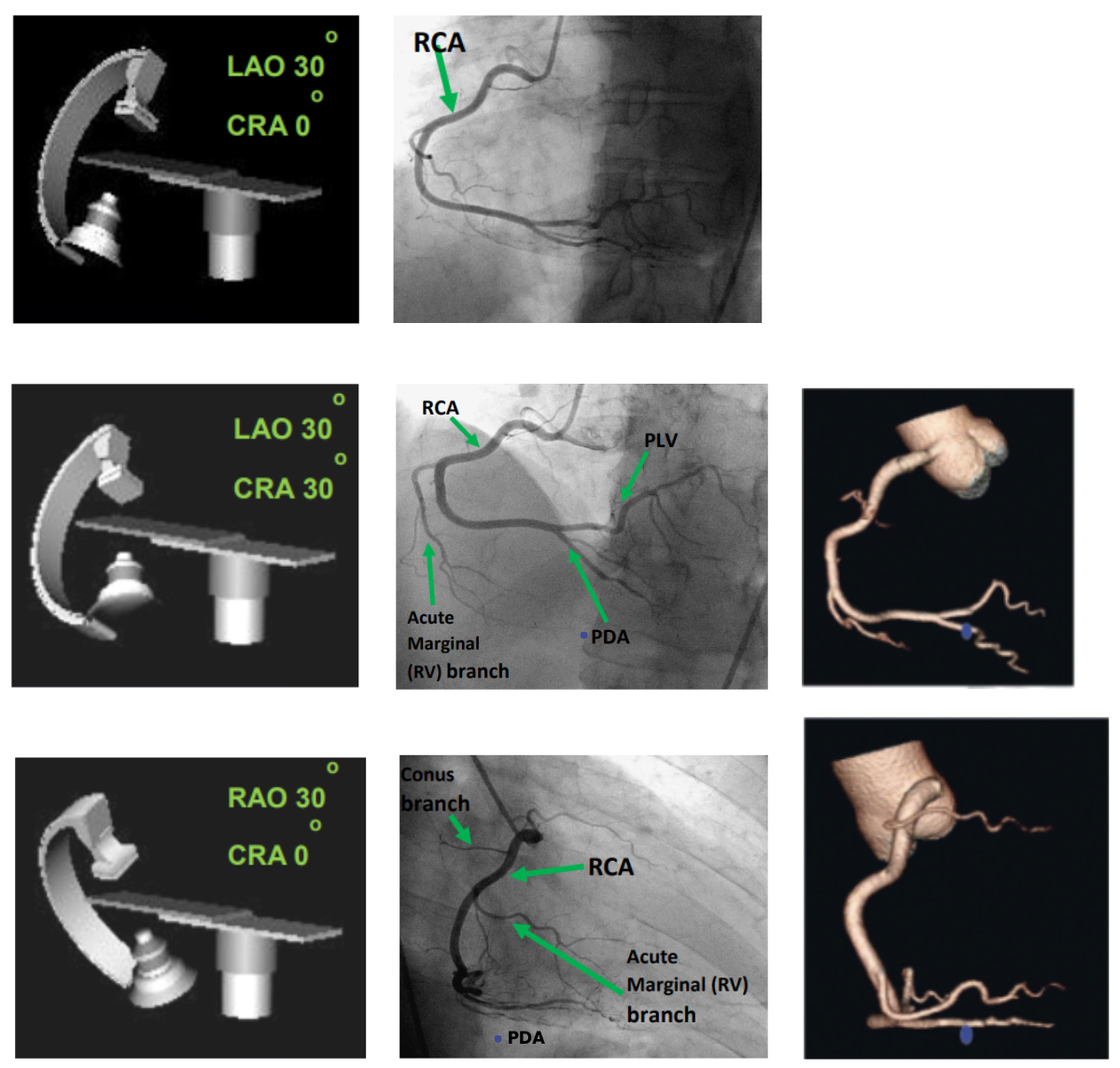
Right Coronary Arteries
Typically, 3 views are used to visualize the RCA.
Conus
Tiny vessel that supplies blood to the infundibulum (conus) of the RV/right ventricular outflow tract
Acute Marginal
Wraps around front of heart, supplying right ventricle.
PLV and PDA
The RCA is simplier to understand, with fewer projections. You may notice a thicker PLV that is branching far to the left, supplying the LV - indicates right-sided dominance or issue with the left side.
- PLV (Posterior Left Ventricular) Top
- PDA (Posterior Descending Artery) Bottom
Cardiac Conduction System
How the heart conducts its own electrical impulses with a specialized network of cells!
Pathway of Electrical Conduction
SA node (right atrium, near the entrance of SVC)
AV node
Bundle of His
Right and left bundle branches
Purkinje fibers
Common Pathology
- Arrythmia (bradycardia, tachycardia and atrial fibrillation)
- Heart block (delay or complete block in the conduction of electrical impulses from the atria to the ventricles)